Brain Injuries Were Blast-Related in Iraq, Afghanistan
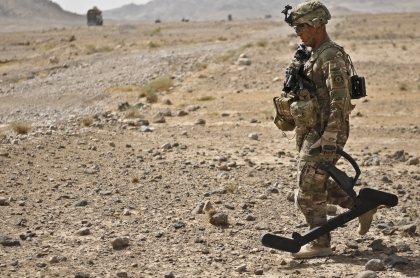
In 2013, an Army sergeant with Engineer Troop, Regimental Support Squadron, Combined Task Force Dragoon, uses a lightweight, portable, handheld device called a Goldie to detect an improvised explosive device (IED) in Kandahar, Afghanistan. Even mild traumatic brain injury caused by the blasts might be linked to development of cognitive issues later in life, according to a new study. U.S. Army Photo by Spc. Joshua Edwards
SEATTLE — Do concussions caused by explosive battlefield blasts increase the risk of developing Alzheimer’s disease (AD)?
A recent study addressed that question and found that alterations in cerebrospinal fluid proteins associated with AD development of Alzheimer’s disease were detected in U.S. veterans deployed to Afghanistan and Iraq who had mild traumatic brain injuries (mTBI) from blasts.
“While our research does not prove that veterans who experienced these injuries will develop Alzheimer’s disease, it raises the possibility that they may be on a pathway leading to dementia,” said first author Ge Li, MD, PhD, an associate professor of psychiatry and behavioral sciences at University of Washington Medicine who works with the Veterans Affairs Northwest Mental Illness Research, Education, and Clinical Center.
The study was led by MIRECC and the Geriatric Research Education and Clinical Center (GRECC) at the VA Puget Sound Health Care System in Seattle.
The recent article in the journal Neurology noted that moderate-to-severe traumatic brain injuries (TBI) have been reported to increase the risk of AD, adding that it is not known whether mild TBI (mTBI) in veterans confers a similar increased risk.1
The study team focused on early AD changes using CSF biomarkers in veterans with blast mTBI. The cross-sectional case-control study of veterans with mTBI and non-mTBI veterans and civilians used two study sources. Blast-mTBI veterans had at least one war zone blast or combined blast/impact mTBI meeting VA and DoD criteria for mTBI. The TBIs were considered mild—and essentially equivalent to a concussion—if the loss of consciousness lasted 30 minutes or less and there was no sign of brain damage on standard clinical MRI or CT scan.
Participants in the concussion group experienced an average of 20 blast concussions and an average of two impact concussions each. Non-mTBI participants had no lifetime history of TBI.
For the study, all participants underwent standardized clinical and neuropsychological assessments and lumbar puncture for collection of the CSF. Researchers said CSF biomarkers were measured using MesoScale Discovery assays for Aβ40 and Aβ42 and INNOTEST ELISAs for phosphorylated tau181 (p-tau181) and total tau (t-tau).
Overall, the sample included 51 participants with mTBI and 85 non-mTBI participants; mean (SD) ages were 34.0 (10.1) and 33.5 years (8.9), respectively. All participants but one were male.
Results indicated that differences in CSF AD biomarkers between mTBI and non-mTBI groups were age-dependent and most obvious in older ages (omnibus test p ≤ 0.08).
“At age 50 years, the mTBI group had lower mean [95% CI] CSF Aβ42 and Aβ40 than the non-mTBI group by 154 [−12 to 319] and 1,864 [610–3,118] pg/mL, respectively,” the authors wrote. “By contrast, CSF p-tau181 and t-tau mean levels remained relatively constant with age in participants with mTBI, while tending to be higher at older ages for the non-mTBI group.”
The study explained that the mTBI group also demonstrated poorer cognitive performance at older ages (omnibus p < 0.08), explaining that “at age 50 years, the mean TMT-B time was higher by 34 seconds [10–58] and the mean CVLT-II short-delay recall was lower by 4.2 points [1.9–6.6]. Poorer verbal memory and verbal fluency performance were associated with lower CSF Aβ42 (p ≤ 0.05) in older participants.”
The team concluded that CSF Aβ levels decreased in middle-age veterans with blast-related mTBI, adding, “These data suggest that chronic neuropathologic processes associated with blast mTBI share properties in common with pathogenic processes known to portend AD onset, thus raising concern that veterans with blast-related mTBI may develop a dementing disorder later in life.”
“Previous research has shown that moderate to severe traumatic brain injuries may increase a person’s risk of Alzheimer’s disease,” explained senior author Elaine Peskind, MD. “What is lesser known is whether mild traumatic brain injuries from military training and combat may also increase a person’s risk. Our study found that these concussions may indeed increase a person’s risk of Alzheimer’s disease.”
The study advised that, with Alzheimer’s disease, the levels of alpha-beta amyloid proteins in spinal fluid typically decrease. Researchers posited that the proteins are deposited in amyloid plaques and remain in the brain instead of being flushed out into the spinal fluid. Tau levels tend to be higher than normal with AD progression, because the proteins are released from dying brain cells, according to the report.
Peskind said she found the decline in beta-amyloid proteins, especially beta-amyloid 42, to be of concern. “A decline in beta-amyloid 42 is the earliest detectable change due to Alzheimer’s that can be found in a cognitively normal person,” she said. “The change can appear as much as 20 years before symptoms. So a person can have the pathology of Alzheimer’s going on in their brain but still not have any symptoms—no problem with their memory or thinking functions—for as long as 20 years.”
The researchers hypothesized that the cause of the changes in the mTBI veterans’ spinal fluid proteins was blast-related damage to the glymphatic system, something that will be examined in a future study.
Memory Tests
All participants took thinking and memory tests. They also had lumbar punctures to collect cerebrospinal fluid. The researchers measured levels of amyloid-beta and tau in the spinal fluid, biomarkers that can be early signs of Alzheimer’s disease.
They found that, with increasing age, those with blast concussion had lower levels of amyloid beta, both Aβ42 and Aβ40, in the spinal fluid than the group without concussion. At age 50, those with blast concussion had Aβ42 levels an average of 154 picograms per milliliter (pg/mL) lower than the group without concussion; Aβ40 levels in those with blast concussion were 1,864 pg/mL lower than the group without concussion. The results were unchanged with the presence of the APOE-ε4 allele, a genetic risk factor for Alzheimer’s disease. The researchers did not find a difference in spinal fluid tau levels between the two groups.
Lower levels of spinal fluid amyloid were associated with poorer performance on memory and thinking tests at older ages, according to the findings. At age 50, for a trail-making test where participants connect a series of dots as quickly as possible while remaining accurate, the average time for the group with concussions was 34 seconds longer than the group without concussions. In addition, on a test that measures verbal memory and includes asking people to recall words after a 20-minute delay, those with concussion scored an average 4.2 points lower than those without concussion; those with concussion scored 8.8 points compared to those without concussion with 13.1 points.
“A reduction in the levels of Aβ42 in the spinal fluid had been shown in other studies to be a marker of amyloid accumulation in the brain, reflecting one of the earliest steps in the development of Alzheimer’s disease,” Peskind pointed out in a press release. “The levels we saw in this study began at around age 45, approximately 20 years earlier than is seen in the general population.”
“Our data show that biomarkers in the spinal fluid associated with concussions from blasts share some properties with the processes that lead to Alzheimer’s disease later in life,” she added. “While our research does not fully address whether veterans who experience these injuries will develop Alzheimer’s disease, it raises the possibility that they may be on a pathway leading to dementia.”
One limitation of the study was that the study group was young and had a small number of participants older than 45, an age earlier than the process underlying Alzheimer’s disease usually begins to emerge. Li said longer studies with more participants are needed that incorporate scans that measure amyloid levels in the brain.
- Li G, Iliff J, Shofer J, Mayer CL, Meabon J, Cook D, Pagulayan KF, Raskind MA, Zetterberg H, Blennow K, Peskind ER. CSF β-Amyloid and Tau Biomarker Changes in Veterans With Mild Traumatic Brain Injury. Neurology. 2024 Apr 9;102(7):e209197. doi: 10.1212/WNL.0000000000209197. Epub 2024 Mar 13. PMID: 38478804.